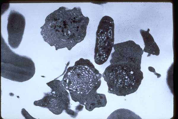
0137094-Abnormal-megathrombocytes-and-platelets-TEM-M-7-leukemia
Abnormal Megathrombocytes and Platelets. Transmission electron micrograph (TEM). Acute Megakaryocytic Leukemia (M-7) in relapse. Buffy coat.
Abnormal Megathrombocytes and Platelets. Transmission electron micrograph (TEM). Acute Megakaryocytic Leukemia (M-7) in relapse. Buffy coat.
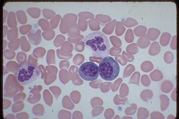
Two Plasmacytoid Lymphocytes, 1 band and 1 mature neutrophil. Side edge of smear can be another site where large, and/or immature, and/or abnormal nucleated cells accumulate. Severe arthritis with osteoporosis blood – 100X
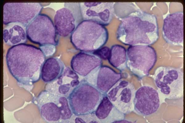
Many Blasts and late neutrophils. Tail area on blood smear. Acute Megakaryocytic Leukemia (M-7) untreated. Blood – 100X
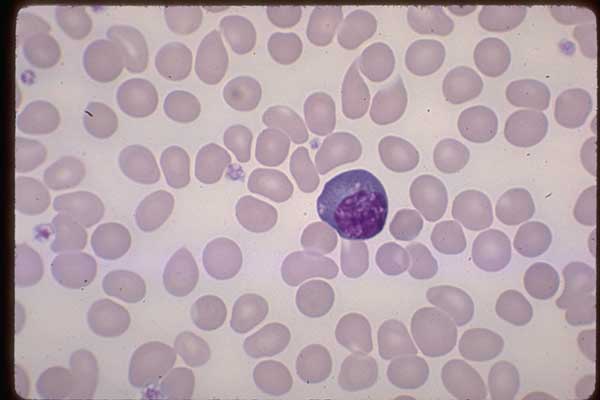
One Plasma Cell containing several vacuoles which represent stored immunoglobulin which does not stain with Wright’s-Giemsa. The red cells lack of central pallor is a preparation artifact seen in thin areas of a smear. The platelets are slightly swollen due to this being an EDTA anticoagulated sample. Alcoholic with liver disease blood – 100X
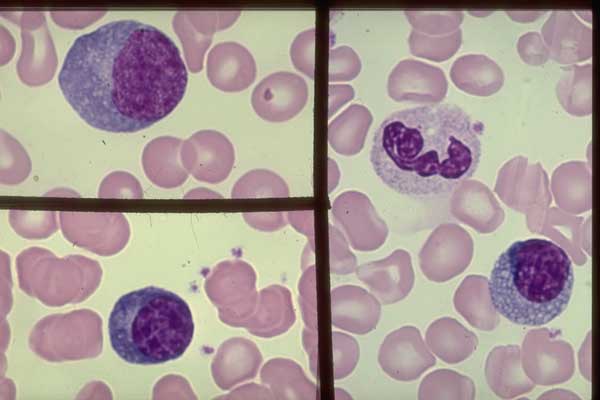
Plasma Cells in each frame. They vary in size but all have an eccentrically located nucleus, blue cytoplasm, a light area (Golgi) adjacent to the nucleus which has a dense chromatin pattern. The plasma cell in the right frame contains multiple small vacuoles. A mature neutrophil is above it. Severe arthritis with osteoporosis blood – 100X